Monday, February 27, 2012


Comparing and Contrasting Replication, Transcription and Translation


Replication
Transcription
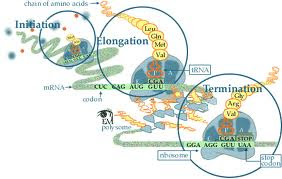
Translation
Initiation

- Helicase unzips the two complementary parent strands


-Single stranded binding proteins anneal to the exposed template strands, preventing them from reannealing


-Primase builds RNA primers which will be used as a starting point by DNA pol III                           


-Transcription factors bind to TATA box

-RNA pol II binds to
 double helical DNA at the promoter region


-DNA strand is unwound exposing the DNA strand

- TFs, TATA box and
 RNA pol II, together
 are called the
initiation complex


- The mRNA, a tRNA with the first amino acid and two ribosomal subunits come together.

-The ribos
omal subunits assemble at the 5'cap of
 the mRNA transcript
sandwiching the mRNA


-Initiation Factors bring the large subunit such that
the initiater tRNA occupies the P site.


- The first tRNA brought into the P site, is carrying methionine because the start codon is AUG
Elongation-DNA Pol III adds nucleotides to the free 3' end of the growing strand using template strand as a guide -leading strand, can be used by polymerases as template for a continuous complimentary strand -The lagging strand is copied away from the fork, discontinuously, in short segment known as Okazaki Fragments RNA pol II begins building the single stranded mRNA in direction of 5' to 3', reading the DNA template in 3' to 5' direction. - The strand of DNA used for transcription is called a template strand. The strand that is not used is known as the coding strand. - This unit is known as the transcription unit.- consists of three step cycles: - codon recognition: in this stage an elongation factor assists the hydrogen bond between the mRNA under A site with the anticodon of tRNA -Peptide bond formation: In this stage an RNA molecule catalyzes the formation of a peptide bond between the polypeptide in the P site with the amino acid in the A site. This step separates the tRNA at the P site from the growing polypeptide chain -Translocation: in this stage the ribosome moves the tRNA with the attached polypeptide from A site to the P site. Translocation ensures that mRNA is "read" 5' to 3' codon by codon
Termination-DNA pol I proofreads the new strand, checking for mistakes. It also replaces the RNA primers with DNAnucleotides -DNA Ligase then "glues" the gaps between the Okazaki fragments. -The mRNA is synthesized till RNA polymerase recognizes the termination sequence at the end of a gene, known as the terminator sequence (AAUAAA) Posttranscriptional Modifications (in eukaryotic cells): - 5' cap is added to the start of the primary transcript -Poly A tail is added to the 3' end - Introns (non-coding parts of the DNA), are taken out by proteins known as spliceosomes ---> Once the primary transcript has been capped and tailed and the intros excised, the process transcript is known as mRNA(messengerRNA) We are now ready to send the mRNA out into the cytoplasm -Termination occurs when one of the stop codons: UAG UGA UAA reaches the A site - A release factor binds to the stop codon and hydrolyzes the bond between the polypeptide and its tRNA in the P site - Now the polypeptide chain is free and the translation complex disassembles.


Replication


Transcription


Translation


Friday, February 10, 2012


Five Remarkable Geneticists

Rosalind Franklin(1920-1957)


Rosalind Franklin made crucial contribution to the solution of the structure of DNA. Franklin is best known for her work on DNA, not only for the excellent X-ray diffraction photographs which she obtained by painstaking and systematic work, but also her insight into what they implied. She also realized that the correct model must have the phosphate groups on the outside of the molecule. Franklin's X-ray diffraction pattern of DNA was crucial evidence for helical structure. Using her photographs Watson and Crick created the famous DNA model. Franklin's photographs are one of the most beautiful X-ray photographs of any substance ever taken.


Arthur Kornberg (1918-2007)


Arthur Kornberg, an American biochemist made outstanding contributions to molecular biology through his research on enzymes. He was the first to isolate DNA polymerase, the enzyme that assembles DNA from its components, and the first to synthesize DNA in a test tube, which earned him the noble prize in 1959. Continuing his work on DNA, Kornberg was eventually able to get DNA polymerase to assemble a 5000-nucleotide DNA chain with the identical form, composition, and genetic activity as DNA from a natural virus. He became the first to replicate an infective virus DNA in vitro.


Frederick Sanger (1918-present)


Frederick Sanger, one of the outstanding biochemists of the modern times found the method for determining the exact sequence of amino acids in proteins and nucleotides in deoxyribonucleic acid (DNA). His methods won him numerous awards, including two Noble prizes in chemistry. Sanger saw a sequence as the key to understanding living matter and set out to determine the exact sequence of amino acids in insulin. He was able to deduce the complete sequence of insulin after twelve years of painstaking research and molecular puzzle solving.


Barbara McClintock (1902-1992)

Leader in the development of maize cytogenetic, McClintock studied chromosomes and how they changed during reproduction in maize. Her groundbreaking work demonstrated many fundamental genetic ideas including genetic recombination by crossing over during meiosis. She produced the first genetic map for the maize linking regions of the chromosomes with physical traits. During 1940s and 1950s, McClintock discovered transposition and used it to show how genes are responsible for turning physical characteristics on and off. In 1983, thirty-five years after publication of her first evidence for transposition, McClintock was awarded the Noble prize for her discovery on mobile genetic elements.


Erwin Chargaff (1905-2002)

To understand DNA scientists were trying to make a model to understand how it works and what it does. In 1940s Erwin Chargaff discovered that DNA is the primary constituent of the gene. He also noticed a pattern in the amounts of the four bases: adenine, guanine, cytosine and thymine. He then took samples of DNA of different cells and found that the amount of adenine was almost equal to the amount of thymine, and that the amount of guanine was almost equal to the amount of cytosine. Thus you can say: A=T and G=C. This discovery later became Chargaff's Rule. Erwin's conclusions helped Watson and Crick in determination of the structure of DNA. His conclusions reasoned because adenine and thymine always exist in the same proportion, so they must always bond together, and similarly for cytosine and guanine. These conclusions lead Watson and Crick winning the Noble prize in 1952.